
Fovea
In the human eye the term fovea (or fovea centralis) is the “pit” in the retina that allows for maximum acuity of vision. The human fovea has a diameter of about 1.0 mm with a high concentration of cone photoreceptors. The centre of the retina, the area of greatest visual acuity. Small depression in the central part of the macula located close to the optical axis of the eye. Central area in the macula that produces the sharpest focus. Contains a high concentration of cones which aid in clear central vision.
Related Links
Fovea of retina — anatomy — Britannica.com
Foveal Avascular Zone ? Webvision
What is the Fovea Centralis? – Definition & Function — Study.com
Fovea – American Academy of Ophthalmology
Structures and functions of the eye: Optic nerve, Fovea & Visual cortex
Related Videos
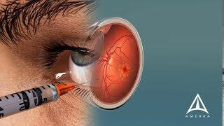
What is Macular Degeneration
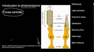
lecture 23 part 2 (Photoreceptors fovea)
Human Eye | aumsum kids education science learn

Fovea centralis
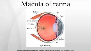
Macula of retina
Photoreceptors (rods vs cones) | Processing the Environment | MCAT | Khan Academy
032 Visual Processing in the Retina
Eye Anatomy: Eyes and Vision Part 1 V2 Newest Version
Macular Degeneration and the Aging Retina

Animation: Dilated Eye Exam
Eye Anatomy and Common Defects Animated Tutorial
The structure of the eye | Processing the Environment | MCAT | Khan Academy
The Human Eye
Central Retina - Part 2 of a Patient's Description of the Retina
2-Minute Neuroscience: The Retina
How the Eye Works Animation - How Do We See Video - Nearsighted Farsighted Human Eye Anatomy
Vision: Crash Course AP 18
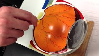
Optic disc Macula lutea fovea centralis rods and cones

What Is The Function Of The Fovea In The Eye
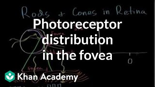
Photoreceptor distribution in the fovea | Processing the Environment | MCAT | Khan Academy
-
What is Macular Degeneration
-
lecture 23 part 2 (Photoreceptors fovea)
-
Human Eye | aumsum kids education science learn
-
Fovea centralis
-
Macula of retina
-
Photoreceptors (rods vs cones) | Processing the Environment | MCAT | Khan Academy
-
032 Visual Processing in the Retina
-
Eye Anatomy: Eyes and Vision Part 1 V2 Newest Version
-
Macular Degeneration and the Aging Retina
-
Animation: Dilated Eye Exam
-
Eye Anatomy and Common Defects Animated Tutorial
-
The structure of the eye | Processing the Environment | MCAT | Khan Academy
-
The Human Eye
-
Central Retina - Part 2 of a Patient's Description of the Retina
-
2-Minute Neuroscience: The Retina
-
How the Eye Works Animation - How Do We See Video - Nearsighted Farsighted Human Eye Anatomy
-
Vision: Crash Course AP 18
-
Optic disc Macula lutea fovea centralis rods and cones
-
What Is The Function Of The Fovea In The Eye
-
Photoreceptor distribution in the fovea | Processing the Environment | MCAT | Khan Academy
What the fovea is and why opticians need to understand it
The fovea is a tiny pit near the center of the retina, the light-sensitive layer at the back of your eye. It is roughly 1.5 millimeters wide. Even though it is small, it is responsible for your sharpest, most detailed vision. When you read a line of text or recognize a face, your fovea is doing most of the work.
As an optician, you will talk about vision clarity every day. Patients ask why their glasses help them see fine print. Employers expect you to explain how lens prescriptions connect to how the eye actually works. Understanding the fovea gives you the foundation to answer those questions clearly and correctly.
The anatomy of the fovea
The retina contains two types of light-sensing cells: rods and cones. Rods handle low-light and peripheral (side) vision. Cones handle color and fine detail. The fovea is packed almost entirely with cones, specifically a type called L-cones and M-cones, which detect red and green wavelengths.
The foveal pit
The fovea has a small depression, called the foveal pit, where other retinal layers thin out and move aside. This arrangement gives incoming light a clear, direct path to the cone cells underneath. Fewer layers between the light and the cones means a sharper image.
The foveola
Inside the fovea sits an even smaller zone called the foveola. It measures about 0.35 millimeters across. The foveola contains only cones and no rods at all. This is the single most precise point of human vision. When you look directly at something, you are pointing your foveola at it.
The surrounding macula
The fovea sits within a larger area called the macula. The macula extends a few millimeters around the fovea and contains a mix of cones and rods. Vision in the macula is still quite good but less sharp than at the foveal center. The yellow pigment that gives the macula its color, called macular pigment, helps filter out harmful blue light and protect the cones underneath.
How the fovea connects to visual acuity
Visual acuity (the clinical term for sharpness of sight, measured on a Snellen chart) depends directly on foveal function. Standard acuity is described as 20/20. This means you can see at 20 feet what a person with typical vision sees at 20 feet. The fovea is what makes 20/20 possible.
When the fovea is healthy and the optical system of the eye, including the cornea and lens, focuses light precisely onto it, vision is sharp. When the fovea is damaged or when the eye’s optics place the focused image in front of or behind the fovea, vision becomes blurred. Prescribing the right corrective lenses moves the focused point back where it belongs: onto the fovea.
Refractive errors and the fovea
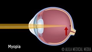
Myopia (nearsightedness) means light focuses in front of the retina, short of the fovea. Hyperopia (farsightedness) means light focuses behind the retina, past the fovea. Astigmatism means light focuses at two different points rather than one clean spot. In every case, the goal of the corrective lens is to redirect light so it converges precisely on the foveal center.
When you work with a prescription, you are working to satisfy one primary requirement: get the clearest possible image onto the fovea. This is why lens power, axis, and cylinder measurements matter so much in opticianry.
Conditions that affect the fovea
As an optician, you are not a diagnostician. Diagnosing eye disease is the role of an optometrist or ophthalmologist. But you do need to recognize when a patient’s symptoms may point to a foveal problem, because that tells you when to refer them to another provider rather than simply adjusting their lenses.
Macular degeneration
Age-related macular degeneration, often called AMD, is one of the most common causes of vision loss in adults over 50 in the United States. AMD damages the macula and, as it progresses, the fovea. Patients with AMD often notice a blurry or dark spot in their central vision. They may have trouble reading or recognizing faces even with their current glasses. No lens prescription corrects AMD. These patients need a referral and often benefit from low-vision aids.
Macular holes
A macular hole is a small break that forms in the foveal tissue. It usually happens due to age-related changes in the vitreous, the gel that fills the inside of the eye. Patients may describe a missing or distorted spot directly in the center of their vision. A macular hole requires surgical evaluation, not a new prescription.
Central serous retinopathy
Central serous retinopathy occurs when fluid builds up under the retina near the macula. It causes the central vision to appear blurry or slightly distorted. It is more common in younger adults, particularly men, and is often linked to stress. Again, this is a referral situation, not a lens adjustment.
Diabetic macular edema
Diabetes can cause blood vessels in the retina to leak fluid into the macula. This is called diabetic macular edema, and it directly threatens foveal function. Patients with diabetes need regular eye exams. As an optician, you may be one of the first people in the eye care chain to notice that a patient’s vision is not improving with new lenses, which is a reason to prompt a referral.
The fovea in low-light and peripheral vision
The fovea works best in good lighting. In dim conditions, your central vision actually becomes less useful. This is why you can sometimes see a faint star better by looking slightly to the side of it rather than directly at it. That technique works because the rod-rich areas surrounding the fovea are more sensitive in low light.
For patients who ask why their night vision seems worse than their daytime vision, this anatomy is part of the explanation. The fovea, built for detail and color, is less active in darkness. Peripheral retinal cells take over. Corrective lenses address the optical focus, but they cannot change the underlying biology of rod and cone distribution.
Why this knowledge matters for the ABO exam
The American Board of Opticianry (ABO) exam tests your knowledge of ocular anatomy as well as optics and lens fitting. The ABO is the national certification body for opticians in the United States. Questions about the retina, the macula, and the fovea appear in the anatomy section of the exam. You are expected to know the difference between the fovea and the foveola, the role of cones versus rods, and how refractive errors shift the focal point relative to the fovea.
You should also know how foveal conditions affect a patient’s functional vision and when a lens prescription can and cannot help. The exam tests whether you can apply anatomy to real patient scenarios, not just recall definitions.
Study tips for ocular anatomy
Start by drawing the layers of the retina from memory. Label the fovea, foveola, and macula on a diagram. Then connect each structure to a clinical situation. Ask yourself: if the fovea is damaged, what does the patient report? If light focuses in front of the fovea, what refractive error is that? Working from structure to clinical outcome helps the material stick.
Use flashcards for the measurement details. The fovea is roughly 1.5 mm wide. The foveola is about 0.35 mm wide. The macula extends a few millimeters around the fovea. These numbers are specific enough to appear in multiple-choice questions.
Communicating foveal function to patients
You do not need to teach patients retinal anatomy. But you do need to explain why their vision works the way it does. The ability to translate clinical facts into plain language is one of the practical skills opticians use every day.
For example, if a patient asks why they see clearly in the center but not the edges of their visual field, you can explain that the center of their vision depends on a specific part of the retina built for fine detail. If they ask why their reading vision has changed with age, you can explain that the lens of the eye stiffens over time, making it harder to focus light precisely onto that central area.
Good communication builds patient trust. It also helps patients understand why their prescription matters and why they should come back for regular exams.
Next steps toward a career in opticianry
Understanding the fovea is one piece of the broader knowledge base you need to pass the ABO exam and work confidently as a certified optician. Anatomy, optics, lens dispensing, and patient communication all connect in this career.
If you are ready to build that knowledge through a structured program, explore the training options available to you. Accredited programs teach you the anatomy, optics, and clinical skills that employers expect. Some states require a license in addition to national certification, so check the requirements for the state where you plan to work.
Browse the program listings on this site to find schools that fit your schedule, location, and budget. Taking that first step toward enrollment is how the preparation begins.