
Choroid
The pigmented layer of the eye between the retina and the sclera. It contains a rich network of blood vessels.
Related Links
Choroid of the eye: MedlinePlus Medical Encyclopedia Image
Choroid – American Academy of Ophthalmology
Choroid – an overview — ScienceDirect Topics
What is the choroid? — Eye and Vision – Sharecare
Choroidal Disorders, Eye Disease. Choroiditis symptoms — Patient
Sclera — White of the Eye – Definition and Detailed Illustration
Anatomy and Structure of the Eye — BrightFocus Foundation
Related Videos
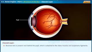
Structure of Eye - Choroid Layer | Class IX Biology
Eye Model.mov
Eyeball Anatomy

Choroid
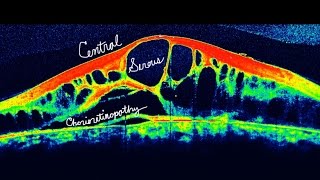
Choroidal Diseases
Part 1 - Detailed Anatomy of the Eye - The Cornea Iris Ciliary body Choroid Explained
Uveal Tract (Choroid Blood Supply) - Anatomy - Part 3/3
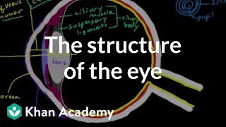
The structure of the eye | Processing the Environment | MCAT | Khan Academy
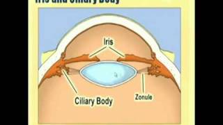
OPTHALMOLOGY -- ANATOMY AND PHYSIOLOGY OF THE EYE
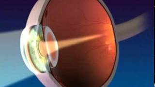
Anatomy and Function of the Eye

Dr. Carol Shields shares new treatment for Choroidal Melanoma via Aura Biosciences

Cataract surgery in iris lens and choroid coloboma.
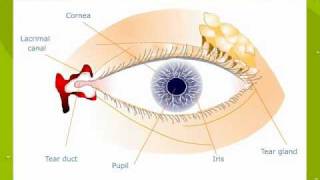
External features of The Human Eye
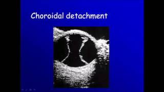
Ophthalmic ultrasonography: 4 choroidal detachment

Lecture A - Eye and Vision
-
Structure of Eye - Choroid Layer | Class IX Biology
-
Eye Model.mov
-
Eyeball Anatomy
-
Choroid
-
Choroidal Diseases
-
Part 1 - Detailed Anatomy of the Eye - The Cornea Iris Ciliary body Choroid Explained
-
Uveal Tract (Choroid Blood Supply) - Anatomy - Part 3/3
-
The structure of the eye | Processing the Environment | MCAT | Khan Academy
-
OPTHALMOLOGY -- ANATOMY AND PHYSIOLOGY OF THE EYE
-
Anatomy and Function of the Eye
-
Dr. Carol Shields shares new treatment for Choroidal Melanoma via Aura Biosciences
-
Cataract surgery in iris lens and choroid coloboma.
-
External features of The Human Eye
-
Ophthalmic ultrasonography: 4 choroidal detachment
-
Lecture A - Eye and Vision
What the choroid is and why it matters for opticians
The choroid is a layer of blood vessels and connective tissue inside the eye. It sits between the retina (the light-sensitive layer at the back of the eye) and the sclera (the white outer wall of the eye). Its main job is to supply oxygen and nutrients to the outer layers of the retina. Without a healthy choroid, the retina cannot function properly, and vision breaks down.
If you are studying for the American Board of Opticianry (ABO) exam or the National Contact Lens Examiners (NCLE) exam, you will see questions about eye anatomy. The choroid is a tested topic. Understanding it clearly helps you answer those questions correctly and serve your patients better in practice.
The structure of the choroid
The choroid is thin, roughly 0.25 mm thick at the back of the eye. It has several distinct layers, each with a specific role.
Bruch’s membrane
Bruch’s membrane is the innermost boundary of the choroid. It separates the choroid from the retinal pigment epithelium (RPE), which is a single layer of cells that supports the retina’s photoreceptors. This membrane acts as a filter, controlling what passes between the choroid and the retina. As people age, Bruch’s membrane can thicken and calcify, which is one factor linked to age-related macular degeneration (AMD).
Choriocapillaris
Just inside Bruch’s membrane is the choriocapillaris, a dense network of small capillaries. These capillaries are unusually wide and have many tiny openings called fenestrations. Those openings allow nutrients to pass easily into Bruch’s membrane and toward the retina. The choriocapillaris supplies the outer third of the retina, including the photoreceptors. The inner two-thirds of the retina get their blood supply from the central retinal artery instead.
Sattler’s layer
Sattler’s layer contains medium-sized blood vessels. It sits directly outside the choriocapillaris and feeds blood into the capillary network.
Haller’s layer
Haller’s layer is the outermost vascular layer of the choroid. It contains the largest blood vessels. These vessels branch inward toward Sattler’s layer and then into the choriocapillaris.
Suprachoroid
The suprachoroid is a thin space between the choroid and the sclera. It contains collagen fibers, smooth muscle cells, and melanocytes (pigment-producing cells). It is also a route that some drug delivery systems now use to reach the back of the eye.
How the choroid functions in the eye
Supplying the retina
The retina is one of the most metabolically active tissues in the body. It burns through oxygen and nutrients at a high rate. The choroid delivers that supply. Because the choriocapillaris is so close to the photoreceptors, oxygen can diffuse across Bruch’s membrane quickly. This efficient delivery system is why the retina can process visual information so rapidly.
Regulating temperature
Light entering the eye generates heat, especially at the macula where light is concentrated. The choroid acts as a cooling system. Its high blood flow rate carries excess heat away from the retina. This protects the photoreceptors from thermal damage.
Controlling intraocular pressure
The choroid contributes to the regulation of intraocular pressure (the fluid pressure inside the eye). Changes in choroidal blood volume affect the overall volume of the eye and, in turn, its pressure. This connection is one reason why conditions affecting choroidal circulation can influence intraocular pressure readings.
Absorbing stray light
Melanocytes in the choroid produce melanin, a dark pigment. This pigment absorbs stray light that passes through the retina without being detected by photoreceptors. Without this absorption, scattered light would degrade image quality. People with less choroidal pigmentation, such as those with albinism, often experience reduced visual acuity and increased sensitivity to bright light for this reason.
Choroidal conditions you should recognize
As an optician, you are not a diagnosing clinician. However, you need to recognize terms that come up in prescriptions, referrals, and patient records. The following conditions involve the choroid directly.
Age-related macular degeneration
AMD is a leading cause of vision loss in adults over 50 in the United States. The dry form of AMD involves the buildup of drusen (small deposits) beneath the retina, at Bruch’s membrane. The wet form involves abnormal blood vessel growth from the choroid into the retina, a process called choroidal neovascularization. These new vessels leak fluid and blood, which distorts and destroys central vision. Patients with wet AMD often need low-vision aids, and fitting those aids is within the optician’s scope.
Central serous chorioretinopathy
Central serous chorioretinopathy (CSC) happens when fluid leaks from the choroid through Bruch’s membrane and the RPE, pooling under the retina. It causes a blister of fluid that distorts central vision. It is more common in young to middle-aged men and is associated with stress and corticosteroid use. Patients may report that straight lines look wavy or that their central vision appears dim or slightly magnified.
Choroidal melanoma
Choroidal melanoma is the most common primary intraocular tumor in adults. It originates from melanocytes in the choroid. It is serious and can spread to the liver if not caught early. Routine dilated eye exams are the main way it is detected. This is one reason optometrists and ophthalmologists stress regular comprehensive exams, and it is a point worth understanding when you talk with patients about why those exams matter.
Choroidal detachment
A choroidal detachment occurs when fluid or blood accumulates in the suprachoroidal space, separating the choroid from the sclera. It can happen after eye surgery or trauma, or as a result of very low intraocular pressure. It is different from a retinal detachment, though both are urgent conditions requiring prompt referral.
Polypoidal choroidal vasculopathy
Polypoidal choroidal vasculopathy (PCV) is a condition where abnormal vessels in the choroid form polyp-like structures that can bleed and leak fluid under the retina. It shares features with wet AMD and is more common in people of Asian and African descent. You may see this term in records for patients using anti-VEGF injections.
The choroid in optician exam preparation
The ABO written exam tests your knowledge of ocular anatomy, including the uvea. The uvea is the middle vascular layer of the eye, and it has three parts: the iris, the ciliary body, and the choroid. Knowing that the choroid is part of the uvea, and understanding each structure’s role, helps you answer anatomy questions with confidence.
Common exam topics related to the choroid include:
- The layers of the eye in order from outermost to innermost
- Which structures make up the uveal tract
- The blood supply to the retina and how it differs between the inner and outer retina
- How AMD and choroidal neovascularization affect vision
- The role of melanin in reducing stray light scatter
When you study eye anatomy, it helps to draw the layers rather than just read them. Place the sclera, choroid, and retina in order, then label the choroid’s sub-layers from outside in: Haller’s layer, Sattler’s layer, choriocapillaris, Bruch’s membrane. Connecting the structure to its function makes the information stick.
How choroidal knowledge applies in daily optician work
You will spend most of your working day fitting frames, adjusting lenses, and explaining prescriptions. Choroidal anatomy does not come up in every conversation. But there are moments when it matters directly.
When a patient hands you a record that mentions “choroidal neovascularization” or “CSC,” you need to understand why their prescription might change more quickly than average. These conditions affect the retina’s shape and function, which in turn affects refraction. A patient with active wet AMD may have a shifting prescription and may need more frequent updates.
When a patient asks why their doctor recommended low-vision devices, knowing that the choroid’s abnormal vessels have damaged their central retina gives you the background to explain it plainly. Patients trust opticians who can speak clearly about their condition, even in simple terms.
When a patient with a high minus (myopic) prescription comes in, note that high myopia is associated with a thinner choroid and higher risk of choroidal complications. This is one reason high myopes need regular dilated exams, not just refraction checks. You can reinforce that message when patients ask whether they really need a full exam.
Next steps for building your optician knowledge
A solid grasp of the choroid is one part of the broader anatomy knowledge required for ABO certification. Anatomy questions appear alongside optics, lens design, and dispensing topics on the exam. The more connected your knowledge is across all those areas, the better prepared you will be on exam day.
Take time to review the full structure of the uvea, then move to the retina and the anterior segment of the eye. Build a clear mental map of each layer and its function before you tackle practice questions. Practice questions will show you which connections you still need to strengthen.
If you are ready to take the next step, explore optician training programs that prepare you for ABO and NCLE certification. A structured program gives you the anatomy, optics, and clinical knowledge you need in a sequence that makes sense, and connects that learning to hands-on dispensing practice.